Multivariate pattern analysis of fMRI data to characterize the cortical representation of pain in humans

Multivariate pattern analysis of fMRI data to characterize the cortical representation of pain in humans

Functional neuroimaging studies have shown that a large array of brain areas is activated when experiencing pain. Yet, how pain emerges in the human brain remains largely unknown. Indeed, painful stimuli are also arousing stimuli that capture attention. Therefore, distinguishing brain activity related to nociception and the perception of pain from brain activity related to stimulus-triggered arousal and attentional capture is challenging. In collaboration with Prof. M. Liang (Tianjin Medical University) and Prof. G.D. Iannetti (University College London and Italian Institute of Technology), Prof. Andre Mouraux and its team from the Institute of Neuroscience (IoNS, http://www.uclouvain.be/ions) have recently been able to isolate, using a multivariate pattern analysis of functional MRI data, features of stimulus-evoked brain activity that distinguishes responses to painful and non-painful stimuli regardless of their intensity and saliency, as well as features that distinguish responses to varying levels of stimulus salience regardless of whether the stimuli generate pain (Liang et al., Cereb Cortex 2019). The identified response...
Read More
Categorical representation from sound and sight in the ventral occipito-temporal cortex of sighted and blind

Categorical representation from sound and sight in the ventral occipito-temporal cortex of sighted and blind

The ventral occipito-temporal cortex (VOTC) is well-known to display topographically organized responses to distinct visual categories (e.g. faces, places, tools etc.). What factors shape this categorical organization? Some have suggested that the main driver is a preference for some specific visual features (e.g. spatial frequencies, eccentricity, curvatures etc.). Others suggested that part of this categorical response cannot be explained by visual features only. With the CPP (Crossmodal Perception and Plasticity, https://cpplab.be/ ) lab, Stefania Mattioni thought to tackle the problem by investigating the categorical response of VOTC in absence of vision; by presenting sounds of different categories in sighted and congenitally blind people.  They reasoned that if the categorical organization of VOTC does not entirely depend on vision, a similar functional profile should emerge when the categories are presented acoustically, even in people without visual experience! They carried out a comprehensive mapping of the representational geometry underlying low-level (acoustic or visual features) and categorical responses to images and sounds in the VOTC of sighted and blind people. They first demonstrated that...
Read More
Towards a landscape of drug-cancer signatures

Towards a landscape of drug-cancer signatures

Large “omics” datasets are increasingly available in public repositories, such as GEO1, ArrayExpress2, Proteome Exchange3 and the GDC4. However, the sheer volume of information requires automated procedures to extract what is biologically relevant for researchers and clinicians. Historically, the NCI60 cell line panel5 (60 cell lines), was the go-to resource that combined phenotypic data with drug response. Later, GDSC6 and CTRP7 have emerged and cover a much greater number of cell lines (987 for GDSC, 860 for CCLE), albeit for a much smaller set of therapies (GDSC1: 320, CTRP: 481) and with a much more incomplete drug-cell line matrix. The next step in the evolution, is the Cancer Dependency Map (https://depmap.org/). The CDM integrates several projects, such as gene expression from CCLE, CRISPR knockouts8, proteomics9 and the PRISM10 drug repositioning screen (578 cell lines, 4686 drugs). Ironically, when it comes to investigating the sensitivity to drug treatment in a cancer-specific context, limitations of the data become immediately apparent. For example, for the average cancer type only 45 cell lines are available. By...
Read More
Cracking the brain code using machine learning

Cracking the brain code using machine learning

At the CPP lab (https://cpplab.be/), headed by Olivier Collignon, the team aims to understand the functional organization of the brain and how different brain networks interact to perform a specific perceptual/cognitive function. Recently, the groundbreaking combination of  functional magnetic resonance imaging (fMRI) -or other neuroimaging methods- with advanced machine learning (ML) or artificial intelligence (AI) techniques has opened unprecedented avenues to understand brain functions in healthy people and patients. The CPP lab uses classification algorithms from ML applied to multivariate neural data to predict the perceptual state of a participant or a patient while perceiving different visual, auditory or tactile stimuli.  Moreover, by combining ML with representational similarity analyses they can understand the format a brain region is using to represent information. Finally, the laboratory is interested in contrasting the representation implemented in deep neural network (DNN) with the one implemented in brain regions to further understand brain organization. They recently applied these techniques to understand how brain networks reorganise in the...
Read More
Applying deep learning tools to improve and automate radiation therapy treatments in oncology

Applying deep learning tools to improve and automate radiation therapy treatments in oncology

The laboratory of Molecular Imaging, Radiotherapy, and Oncology (MIRO) works on a variety of projects that combine technical and clinical knowledge with the aim of improving radiation oncology treatments. Radiation therapy is one of the most used modalities to treat cancer and it uses different types of medical images, like computed tomography (CT), positron emission tomography (PET), or magnetic resonance images (MRI), for diagnosis, treatment planning, treatment delivery, treatment verification, and follow-up. Joining the recent rise of artificial intelligence methods applied to the medical domain, the MIRO laboratory is exploring different ways of applying deep learning tools to improve and automate radiation therapy treatments. Meet the members of the MIRO team and read about their success stories! Umair Javaid and Kevin Souris work together to mitigate inherent noise in Monte Carlo dose distributions using deep learning[1]. In particular, they use a U‐Net architecture with dilated convolution kernels (U7 in Figure 1) to denoise dose distributions for proton therapy treatments calculated with a Monte Carlo dose engine [2]. Denoising of dose maps has two advantages: first, it...
Read More
Deep Learning in Nuclear medicine: automatic estimation of thyroid volume

Deep Learning in Nuclear medicine: automatic estimation of thyroid volume

In nuclear Medicine, radioactive compounds are administered to patients in order to get both functional and anatomical data. With Anger Cameras and specific cristals, one can collect an image from the distribution of the tracer, which is called a “scintigraphy”. In thyroid scintigraphy, several pathological situations may occur. François-Xavier Hanin, nuclear medicine physician (CHU UCL Namur) developed a deep learning model based on a U-net architecture to automatically analyze these images. First, a segmentation system was developed to define the contours of the thyroid. This has potential applications in automatic assessment of the uptake, a computation of the percentage of injected activity which is absorbed by the thyroid. He then used a similar architecture to detect cold nodules. These nodules have a particular interest in thyroid pathology, as many differentiated thyroid cancers appear as cold nodules (meaning, with no uptake). The U-net Architecture was described in 2015 (1) and its implementation was applied with 6 levels, a maximum of 1024 filter dimension at center, 2 convolutions on the way...
Read More
Voxel-based MRI morphometric analysis program for the detection of cortical abnormalities in pharmacoresistant epilepsy

Voxel-based MRI morphometric analysis program for the detection of cortical abnormalities in pharmacoresistant epilepsy

Epilepsy is one of the most frequent chronic neurological disorders. In 30 to 40% of the patients, seizures cannot be controlled adequately with medication, whose cases are called drug-resistant epilepsy. For 10 to 50% of these patients, surgery constitutes a valuable treatment option. Aims of the surgery is to control seizures by resection of the epileptogenic tissue without causing damage to areas of the brain responsible for essential functions. Candidates for lesionectomy undergo an extensive pre-surgery evaluation. Magnetic resonance imaging (MRI) is of paramount importance in this presurgical evaluation to detect and delineate lesions. Detection of a previously undetected lesion can drastically change the presurgical planning and significantly improve seizure post-surgery outcome. Yet some kind of lesions, as subtle focal cortical dysplasia (FCD), are more difficult to detect because of their small sizes, and often buried in the complex convexities of the cortex. Practical constraints of time may lead MRI readers to miss those lesions that require increased scrutiny. In a prospective trial, Professor Riem El Tahry & Professor Susana Ferrao santos (Center for...
Read More
Automated, fast and reliable analysis of pyrosequencing signals

Automated, fast and reliable analysis of pyrosequencing signals

Pyrosequencing is a DNA sequencing technology that has many applications including rapid genotyping of single nucleotide polymorphisms (SNP) and other sequence variations between different cells, individuals and species. Identifying the specific composition of microorganisms inside a microbial population may help choose the most specific treatment in case of infectious disease. Likewise, pyro-sequencing can also apply to the screening and identification of oncogenic mutations(s) in a heterogeneous sample containing tumour cells of interest in a background of wild type cells. Converting a pyrosequencing signal into a nucleotide sequence appears highly challenging when signal intensities are low (unitary peak heights <5) or when complex signals are produced by several target amplicons. In these cases, the pyrosequencing software fails to provide correct nucleotide sequences. At the CTMA (Center for Applied Molecular Technologies), researchers developed the AdvISER-PYRO, an algorithm based on a machine learning method (i.e., a sparse representation) to perform an automated, fast and reliable analysis of pyrosequencing signals which circumvents above...
Read More
A new microstructure fingerprinting approach for quantitative microstructure estimation of the brain white matter

A new microstructure fingerprinting approach for quantitative microstructure estimation of the brain white matter

Because it is sensitive to the microscopic displacements of water molecules diffusing in the tissues, diffusion-weighted magnetic resonance imaging (DW-MRI) has become the modality of choice to study the anatomy of the white matter at the micrometer level, with applications in the diagnosis and treatment of many neurological disorders and brain injuries. Our new approach at the intersection of physical modeling, numerical simulations and signal processing proposes a way to bridge the gap between the native millimeter resolution of MRI and neuroanatomical indices at the micrometer scale. We use specific MRI acquisitions known as diffusion-weighted MRI which by design are sensitive to the micrometer-scale diffusing motion of water molecules in the tissues at body temperature. In traditional approaches, forward mathematical models based on analytical formulas are devised to link the geometry of white matter tissue, the diffusion of water molecules in that geometric arrangement and the physics of MRI acquisitions. The free parameters of such a forward model are then adjusted...
Read More
Proton Radiography to reduce range uncertainty in therapy

Proton Radiography to reduce range uncertainty in therapy

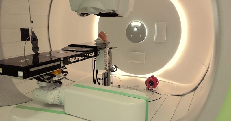
ImagX.R, the research group on image guided proton therapy, has the pleasure to announce the PhD defence of Sylvain Deffet. He will present his research on proton radiography and discuss how this method could improve treatment outcome in proton therapy. Proton therapy is an advanced form of radiation therapy which is increasingly used worldwide. Unlike photons, protons deliver a sharp dose at a precise location corresponding to their range in the patient. Thanks to this physical property, proton therapy has the potential to spare healthy tissues better than conventional radiation therapy. However, a consequence of this dosimetric property is that the range of the protons inside the patient must be accurately predicted to deliver the dose as planned. Unfortunately, several uncertainties arising during treatment planning may significantly impact the range of the protons and hence jeopardize dose conformity. To better quantify and potentially reduce the uncertainties, the implementation of imaging techniques that would provide a direct information on the energy reduction...
Read More