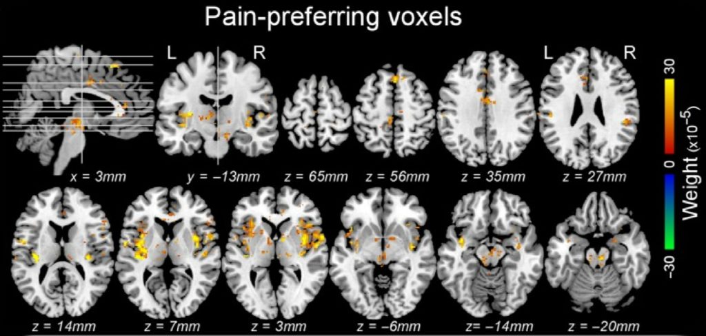
Functional neuroimaging studies have shown that a large array of brain areas is activated when experiencing pain. Yet, how pain emerges in the human brain remains largely unknown. Indeed, painful stimuli are also arousing stimuli that capture attention. Therefore, distinguishing brain activity related to nociception and the perception of pain from brain activity related to stimulus-triggered arousal and attentional capture is challenging. In collaboration with Prof. M. Liang (Tianjin Medical University) and Prof. G.D. Iannetti (University College London and Italian Institute of Technology), Prof. Andre Mouraux and its team from the Institute of Neuroscience (IoNS, http://www.uclouvain.be/ions) have recently been able to isolate, using a multivariate pattern analysis of functional MRI data, features of stimulus-evoked brain activity that distinguishes responses to painful and non-painful stimuli regardless of their intensity and saliency, as well as features that distinguish responses to varying levels of stimulus salience regardless of whether the stimuli generate pain (Liang et al., Cereb Cortex 2019). The identified response features were spatially distributed and could not be ascribed to a specific brain structure, compatible with the view that pain may emerge from neural activity occurring within a distributed large-scale brain network.

References :
Liang M, Su Q, Mouraux A, Iannetti GD. Spatial Patterns of Brain Activity Preferentially Reflecting Transient Pain and Stimulus Intensity. Cereb Cortex. 2019;29(5):2211–2227.
Su Q, Qin W, Yang QQ, et al. Brain regions preferentially responding to transient and iso-intense painful or tactile stimuli. Neuroimage. 2019;192:52–65.
Liang M, Mouraux A, Hu L, Iannetti GD. Primary sensory cortices contain distinguishable spatial patterns of activity for each sense. Nat Commun. 2013;4:1979.
